
Cryostats
The cryostat is an essential tool in dermatology, aiding in the accurate diagnosis and treatment of various skin disorders and supporting ongoing research in the field.
Here's how a dermatology office might utilize a cryostat:
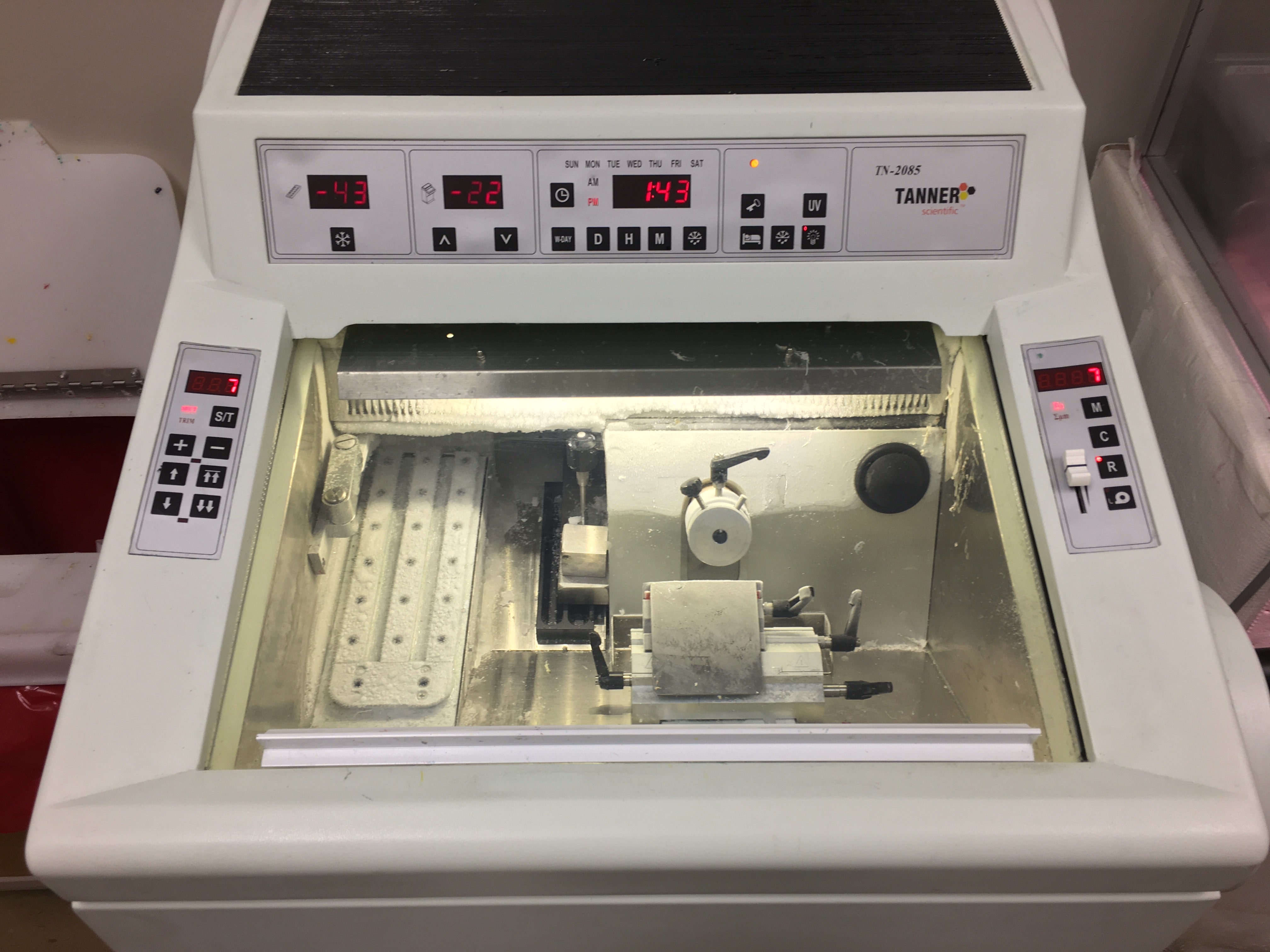
A Cryostat is used to cut very thin sections of frozen tissue, typically at temperatures below freezing. In dermatology, this allows the dermatologist or pathologist to examine the tissue samples at a microscopic level to identify skin diseases, such as skin cancer, melanoma, and other dermatological conditions.
Dermatologists use the Cryostat to prepare tissue samples taken from skin biopsies for analysis. This analysis helps in the diagnosis of various skin conditions, including but not limited to skin cancer, infectious diseases, autoimmune disorders, and inflammatory skin conditions.
In some cases, dermatologists may use the Cryostat for immunofluorescence studies, which help in diagnosing autoimmune skin diseases. The Cryostat allows for the preparation of thin sections of tissue that can be examined under a fluorescence microscope to detect the presence of specific antibodies or antigens.
Dermatology offices and research facilities use Cryostats to support ongoing research and development in the field of dermatology. This may involve studying the effects of various treatments on skin tissue or investigating the underlying mechanisms of certain skin conditions.
Cryostats may also be used in dermatology offices for educational purposes, allowing medical students, residents, and fellows to learn about the preparation and examination of skin tissue samples.
Any questions?
If we have questions about your cryostat please contact us or submit a service request below.